
Echocardiography · Sibiu
Echocardiography in Sibiu, with the liver, carotid arteries and thyroid checked in the same session
A heart ultrasound shows how your heart looks and how it works. In the same session we take a preliminary look at the liver, the carotid arteries and, when needed, the thyroid, at no separate cost.
What echocardiography is and what it shows about your heart
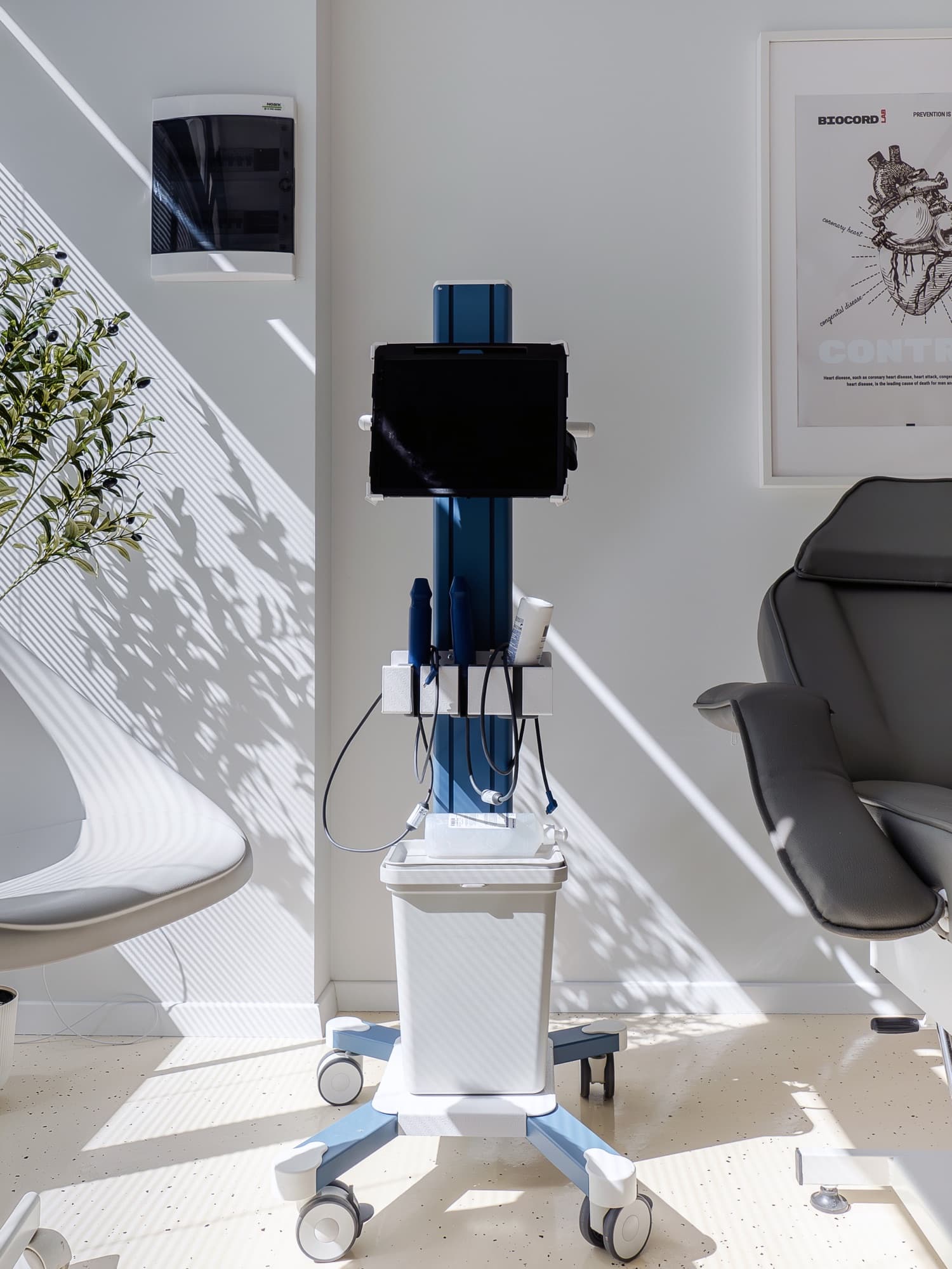
Echocardiography, also known as a heart ultrasound, is an ultrasound scan that shows the heart moving, in real time. You can see how it contracts, how it relaxes, how the valves open and close, and how the blood flows through them.
It is a non invasive test. No radiation, no injections and no pain. A probe glides over the skin of your chest with a little gel, and the image appears on the screen while the examination is happening.
It is not the same thing as an electrocardiogram. That one shows the electrical activity of the heart, meaning the rhythm. Echocardiography shows structure and function, meaning the walls, the valves and the pumping power. They are two different questions, and a full answer often needs both.
At the Biocord clinic, the heart ultrasound does not stop at the heart. In the same session we take a preliminary look at the liver, for signs of fatty change, at the carotid arteries, for atherosclerotic plaque, and, when your symptoms call for it, at the thyroid. All of this is part of the echocardiogram and is not charged separately, and the results go into your cardiometabolic picture rather than staying isolated observations.
“Preliminary” means exactly what it says. The assessment shows whether there is something worth following or investigating further. It does not replace a full vascular Doppler scan or an endocrinology consultation, and when you need one of those we tell you clearly and explain why.
When an echocardiogram is indicated
Some indications start from a symptom, others from an abnormal result or from a diagnosis already made that needs following over time.
- You get short of breath on exertion or when you lie flat
- You tire quickly doing things you used to do without any trouble
- A heart murmur was heard and has never been investigated
- You have had high blood pressure for several years
- You have palpitations or a known rhythm disorder
- Your ankles have swollen or you have gained weight quickly from fluid
- You have a diagnosed valve problem that needs following over time
- You have had a heart attack, or there is heart disease in your family at a young age
- You have fatty liver, prediabetes, metabolic syndrome or high cholesterol
- You smoke, have diabetes, or have relatives who had a heart attack or stroke at a young age, and you want to know whether plaque has formed
- You are about to start a treatment that requires an assessment of heart function
If right now you have chest pain that will not go away, severe difficulty breathing, or you have fainted, do not wait for an appointment. Call 112.
How the examination goes
You do not need any complicated preparation, and you leave with the explanation, not just with the images.
The short conversation beforehand
You tell me what symptoms you have, what treatments you are on and what tests you have already had. Without that context, the image stays just an image.
Getting into position
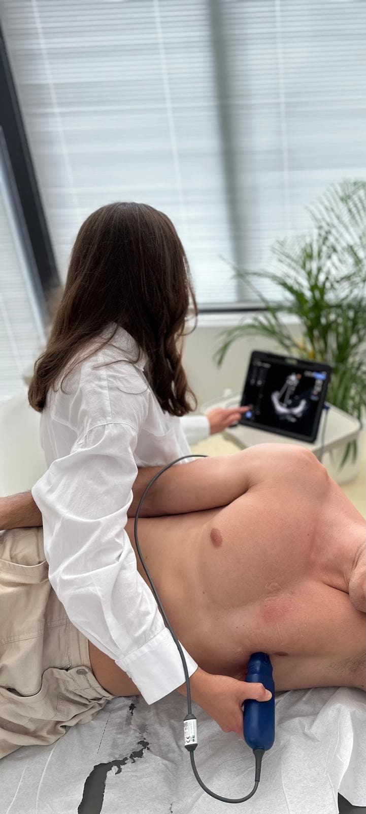
You lie down on the couch, usually on your left side, with your chest uncovered. Gel is applied, which can feel slightly cold, and a few electrodes are attached to follow your rhythm during the examination.
The scan itself
The probe moves over your chest, from several angles. Now and then I will ask you to hold your breath for a few seconds, for clearer images. It does not hurt, at most you feel some pressure between the ribs.
The liver, the carotids and the thyroid
In the same session we take a preliminary look at the liver, for signs of fatty liver, and at the arteries in your neck, for atherosclerotic plaque. When your symptoms call for it, we also look at the thyroid. These are natural parts of a cardiometabolic assessment, included in the echocardiogram, not tests charged separately.
The explanation and the conclusion
I tell you what I saw, in words you can repeat at home. What is normal, what needs watching, what changes in your treatment, and when it is worth repeating the scan.
What an echocardiogram actually shows
The first thing we look at is the pumping function of the left ventricle, expressed as the ejection fraction. It says what proportion of the blood in the ventricle is sent onwards with each beat. When it drops, the picture of heart failure appears, and treatment is built around that figure.
Then the thickness of the walls and the size of the chambers are measured. A thickened wall is the usual sign of blood pressure that has been left too high for too long, and a dilated chamber speaks of an old overload. Both show up before symptoms do, which is what makes them useful in prevention.
The valves are looked at in two ways: how they look and how they let the blood through. This is where the Doppler examination comes in, measuring the speed and direction of flow and showing whether a valve has narrowed, whether it lets blood leak back, and how significant that is.
We also assess the way the heart relaxes between beats, the estimated pressures in the pulmonary circulation, and whether there is fluid around the heart. Put together, all of this often explains a symptom that until then seemed to have no cause.
Fatty liver disease, the metabolic link we look for in the same session
Hepatic steatosis, that is fatty liver, means a build up of fat inside the liver cells. For a long time it was seen as a gastroenterology problem. Today we know that, in its metabolic form, it goes hand in hand with insulin resistance, with metabolic syndrome and with a higher cardiovascular risk.
Most of the time it causes no symptoms at all. It is found by chance, on a scan done for another reason, or it is suspected after slightly raised transaminases. That is exactly why it makes sense to look for it in someone who is coming in for their heart anyway.
On ultrasound, a liver with fat in it looks brighter than normal, and the appearance is described in rough grades, from a mild fat load up to a significant one, where the deeper structures become harder and harder to see. It is a visual judgement, useful as a signal, not an exact measurement.
It is important to be honest about what ultrasound can and cannot do. It sees the fat, but it does not say how much the liver has suffered, meaning it does not assess fibrosis. There are dedicated tests for that, such as elastography. If your results raise that kind of suspicion, I tell you clearly what comes next and where it is done.
The good news is that metabolic fatty liver disease is not a life sentence. It is one of the few situations where the liver can recover, over time, if the cause behind it changes. That is why we record it and follow it, rather than mentioning it in passing at the end of a report.
What actually changes if you have fatty liver
The strongest lever is still weight loss, done gradually. A modest but sustained reduction usually shows in the amount of fat in the liver, and when the loss is larger the rest of the metabolic picture improves as well. Consistency matters more than speed.
When it comes to food there is no magic list of things to avoid, but a few make the biggest difference: sweetened drinks and fruit juices, alcohol, ultra processed products, and large portions of sweets and refined starches. Those load the liver most directly, and cutting them back is something you feel.
On the other side of the scales are vegetables, pulses, fish, olive oil, nuts and wholegrains, in other words the Mediterranean pattern. It is not a diet you keep for three weeks, it is a way of eating you can carry on with for years.
Exercise helps even without a dramatic drop on the scales. Brisk walking done consistently, plus strength training, build muscle mass and improve the way the body uses insulin. Not enough sleep and untreated sleep apnoea pull in the opposite direction.
Alongside that, we treat the blood pressure, the cholesterol and the blood sugar. Not because it cures the liver, but because in someone with fatty liver disease the first danger is still the cardiovascular one.
The carotid arteries, a window onto atherosclerosis that has no symptoms yet
The carotid arteries are the large vessels on the sides of your neck that carry blood to the brain. Because they sit close to the skin, ultrasound shows them well, and the state of their walls says a lot about what is happening across the whole arterial system.
As part of the echocardiogram we take a preliminary look at them, with a precise aim: to see whether the wall has thickened or whether atherosclerotic plaque has formed. In other words, atherosclerosis that already exists but has not yet caused any symptom and does not show up in any blood test.
It matters because atherosclerosis does not confine itself to one vessel. A plaque seen in the neck can move you into a higher risk category than the usual calculation suggests, and where your risk sits decides how firmly cholesterol and blood pressure are treated.
Finding a plaque does not mean surgery, and it does not mean an event is coming. It usually means stricter targets for cholesterol and blood pressure and closer follow up. If the appearance raises the suspicion of a significant narrowing, you get a recommendation for a dedicated vascular Doppler scan, which measures exactly how narrow it is.
The thyroid, when heart symptoms start somewhere else
Thyroid hormones act directly on the heart. An overactive thyroid speeds up the rhythm and encourages palpitations and atrial fibrillation. An underactive one slows the rhythm, raises cholesterol and brings fatigue and fluid retention. Many symptoms that look cardiac actually start at the thyroid.
That is why, when your picture calls for it, we also take a preliminary look at the thyroid in the same session. The assessment looks for nodules, changes suggesting thyroiditis, or anything else that needs a dedicated endocrinology assessment.
Biocord is a cardiology clinic, not an endocrinology one. The assessment here does not make a thyroid diagnosis and does not replace blood tests, because ultrasound shows what the gland looks like, not how much hormone it makes. Fine needle biopsy and elastography are not done at the clinic. If something needs clearing up, you get a clear recommendation to see an endocrinologist.
Prices
| Service | Price |
|---|---|
| Cardiology consultation + ECG | 400 lei |
| Cardiology consultation + ECG + echocardiography | 620 lei |
| Cardiology follow-up + ECG | 300 lei |
| Echocardiographyincludes an orientative assessment of the liver, the carotid arteries and the thyroid | 400 lei |
| Holter ECG monitoring24 hours | 370 lei |
| Ambulatory blood pressure monitoring (ABPM)24 hours | 370 lei |
| Exercise stress test on a cycle ergometer | 450 lei |
| Body composition analysis | 100 lei |
| Ankle-brachial index (ABI)with interpretation | 100 lei |
| Electrocardiogram (ECG) | 100 lei |
| ECG + SDS (sudden death screening)extended BTL assessment, for people up to the age of 35 | 150 lei |
| Intravenous Ferinject 500 mgmedication and medical supervision included | 800 lei |
| Intravenous vitamin C (Pascorbin 7.5 g)product and medical supervision included | 350 lei |
You have seen the prices. Shall we go ahead?
Tell us what brings you to a cardiologist and we will suggest a time that works for you.
Frequently asked questions
Does an echocardiogram hurt, or involve radiation?
What is the difference between an echocardiogram and an electrocardiogram?
What does transthoracic Doppler echocardiography mean?
Do I need to come on an empty stomach?
What do normal values mean on an echocardiogram?
Does an echocardiogram show whether the arteries of my heart are blocked?
Is the carotid and thyroid check charged separately?
Does it replace a full carotid Doppler scan?
If a thyroid nodule is found, what happens next?
What is a stress echocardiogram?
Do I need a referral letter?
Why does a cardiologist look at the liver as well?
How long does an echocardiogram take and when do I get the result?
Echocardiography in Sibiu
The Biocord practice is at Strada Ion Agârbiceanu 11, Sibiu, in the Turnisor neighbourhood.
Phone: 0771 193 295
Email: bio.cord@yahoo.com
Appointments are made by phone or through the form on this site. We confirm the time before you come.
Open in Google Maps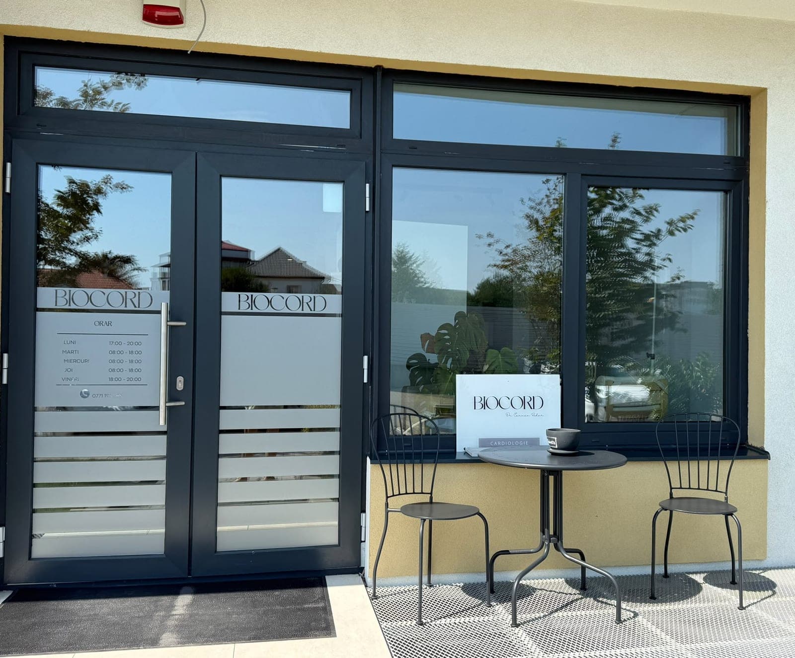
Services that go together with this one
- Cardiology consultationA consultation that does not stop at the reason you came in. We go through your history, your test results, your lifestyle and your risk factors, then build a long term prevention plan together.
- Electrocardiogram (ECG)The basic test of the heart: a few minutes, with no discomfort. Its value lies in the reading, that is, in what your tracing means alongside the rest of your story.
- Body composition analysisWeight on its own says almost nothing. Body composition shows the balance between fat and muscle, and that is where the practical recommendations start.

Book echocardiography
We confirm the time by phone and tell you whether any preparation is needed.