
ECG · Sibiu
An electrocardiogram in Sibiu, read in your context rather than on its own

The basic test of the heart: a few minutes, with no discomfort. Its value lies in the reading, that is, in what your tracing means alongside the rest of your story.
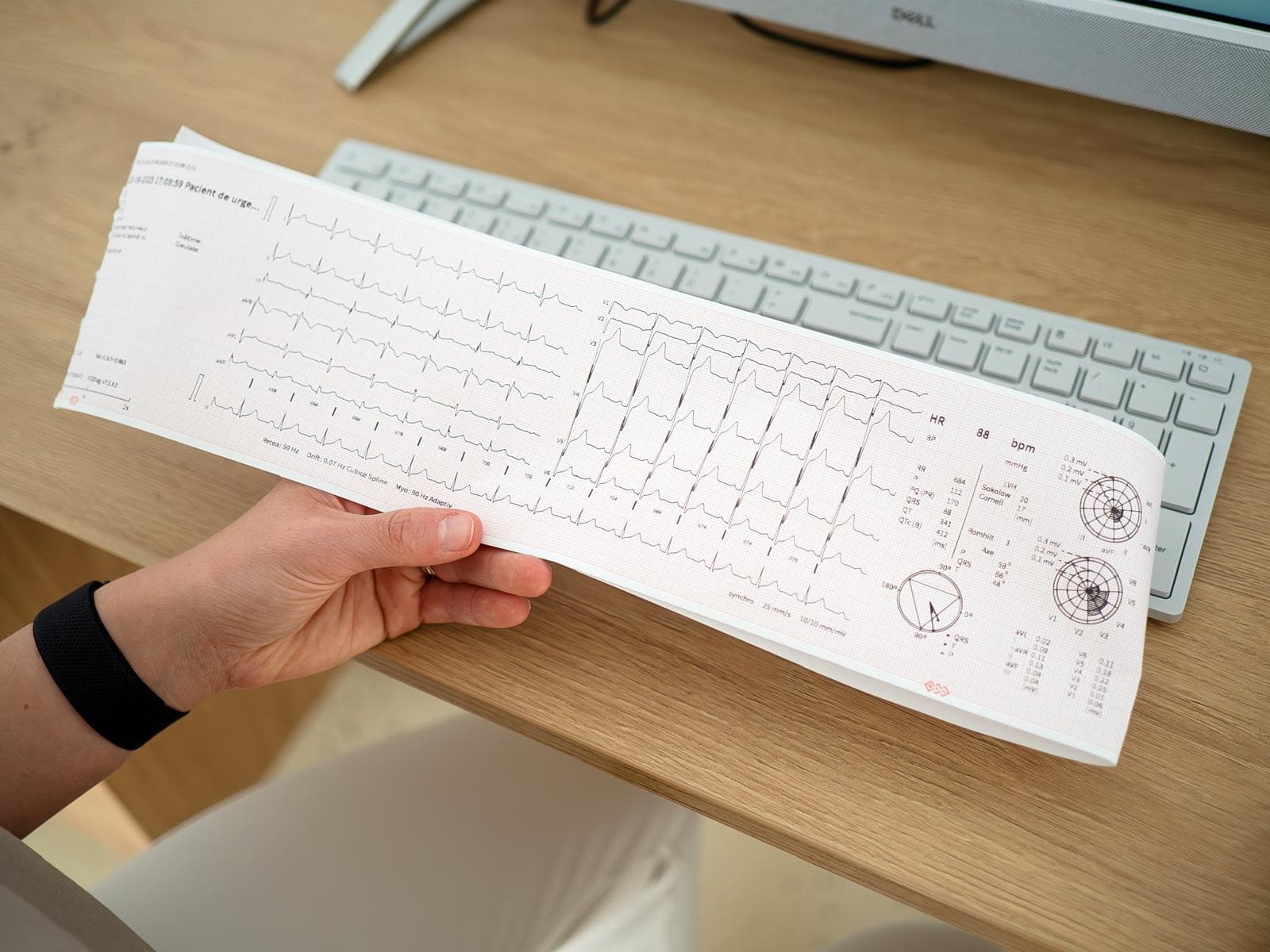
What an electrocardiogram is and what it shows
Every heartbeat starts from an electrical impulse that travels through the heart muscle in a set order. An electrocardiogram records that current from the surface of the skin and draws it as a trace of waves and lines.
Electrodes are stuck to your chest, your arms and your legs, and the machine reads the signal from several directions at once. You feel no current and it does not hurt.
The trace gives us your heart rhythm and rate, rhythm and conduction disturbances, signs of strain from a lack of oxygen, and clues about thickening of the walls. It is a quick, non invasive investigation and an indispensable one in diagnosing and monitoring the health of your heart.
At the Biocord clinic, an electrocardiogram is not done in isolation, but as part of the cardiology assessment. A trace has value when it is read alongside your symptoms, your blood pressure and your echocardiogram. The same picture can mean nothing in one person and change the plan in another.
Worth saying from the start: an electrocardiogram captures a few seconds from the electrical life of your heart. If your symptoms turn up rarely, or only on exertion, a normal trace does not rule them out.
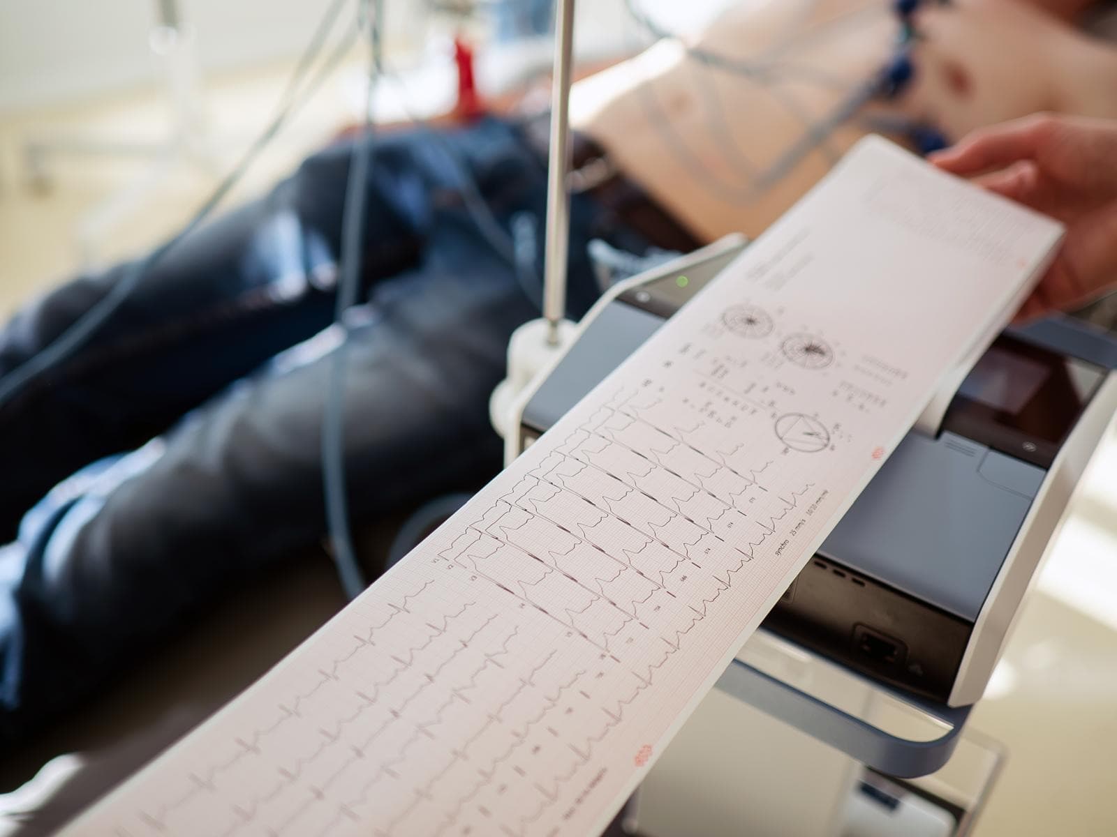
When an electrocardiogram is done
It is the starting investigation in almost any cardiology assessment.
- You feel palpitations, irregular beats, or episodes where your heart seems to be racing
- You have pain or pressure in your chest, especially if it comes on with exertion and eases with rest
- You get tired or run out of breath doing things that used to be easy
- You have dizziness, a feeling that you might faint, or you have fainted
- You have high blood pressure, already diagnosed or newly discovered
- You are starting or adjusting a treatment that can affect your heart rhythm
- You have diabetes, high cholesterol, metabolic syndrome, or you are a smoker
- You have close relatives with heart disease, arrhythmias or sudden death at a young age
- You have no symptoms, but you want a starting point for future comparisons
If right now you have chest pain that will not go away, serious difficulty breathing, cold sweats, or you have fainted, do not wait for an appointment. Call 112.
How it is done, step by step
One of the simplest investigations in the clinic. It takes a few minutes and asks for no preparation.
Getting ready
You lie down on the couch and uncover your chest, your ankles and your wrists. There is no need to come on an empty stomach.
Placing the electrodes
Self adhesive electrodes are applied in fixed positions on your chest and one on each limb. Where they go matters, because the trace is read in relation to those points.
The recording
You lie still, relaxed and without moving, breathing normally and not talking. It takes a few tens of seconds. Cold, shivering or movement can add noise to the trace.
The interpretation
The trace is read straight away, in context: rhythm, rate, axis, intervals and the shape of the waves, compared with what we know about you and with your older electrocardiograms.
What comes next
If everything is in order, the trace stays as a reference point. If changes show up, the next steps are decided: echocardiography, Holter ECG monitoring, 24 hour blood pressure monitoring or blood tests.
The terms that come up most often on an ECG report, in plain language
Plenty of people go home with a piece of paper full of phrases that sound alarming, and nobody to translate them. Below are some of the most common ones. None of them is a diagnosis and none of them can be interpreted outside your own context.
“Sinus rhythm” means the impulse starts where it should, from the heart’s own natural pacemaker, and it describes the normal rhythm. “Sinus tachycardia” is the same rhythm, only fast, and “sinus bradycardia” is the same rhythm, only slow. Both can be physiological: a fever or exertion speeds the heart up, and in trained athletes a low resting rate is common.
“Right bundle branch block” describes a delay in the impulse travelling to the right ventricle. It sounds serious because of the word block, but the incomplete form is also found in people with a structurally normal heart. What matters is whether it appears on its own, whether it is new compared with an older trace, and what the echocardiogram shows. Left bundle branch block is generally looked at with more attention.
“Ectopic beats”, also called premature beats, are extra beats that arrive earlier than they should. They are common and they turn up in healthy people too, on a background of tiredness, coffee, alcohol or stress. They are assessed by how frequent they are and by what the heart looks like on echocardiography.
“Atrial fibrillation” is a rhythm disorder in which the atria stop contracting in an orderly way and the pulse becomes completely irregular. It is one of the most important ones to identify, because it changes the treatment.
“ST-T changes” refer to the part of the trace that describes the electrical recovery of the ventricles. They can signal strain from a lack of oxygen, but also a potassium imbalance or the effect of certain medicines. Here, the comparison with an earlier trace is worth an enormous amount.
“Left ventricular hypertrophy” suggests a thickened wall, most often after blood pressure that has stayed high for too long, and the suspicion is settled with echocardiography. “Axis deviation” only describes the direction the current travels through the heart and, on its own, it says very little.
If you have been given a result with terms you do not understand, bring the paper to the consultation, along with your older traces. The most useful question is not “what does this say”, it is “what does this mean for me”.
What an electrocardiogram cannot show
A normal trace does not guarantee a heart without problems, and that is the most important thing to remember. The recording lasts a few seconds, and an arrhythmia that appears once a week has every chance of being absent at exactly that moment. That is what the Holter ECG is for.
It also does not show what the heart looks like. Wall thickness, chamber size, how the valves work and pumping power are seen on echocardiography. It does not tell you whether there is atherosclerotic plaque either, and for that we look at the carotid arteries during the ultrasound.
On top of that, a trace recorded at rest can be normal in someone whose symptoms appear only on exertion. That is why your story matters: when the symptoms come, what sets them off, how long they last.
Prices
| Service | Price |
|---|---|
| Cardiology consultation + ECG | 400 lei |
| Cardiology consultation + ECG + echocardiography | 620 lei |
| Cardiology follow-up + ECG | 300 lei |
| Echocardiographyincludes an orientative assessment of the liver, the carotid arteries and the thyroid | 400 lei |
| Holter ECG monitoring24 hours | 370 lei |
| Ambulatory blood pressure monitoring (ABPM)24 hours | 370 lei |
| Exercise stress test on a cycle ergometer | 450 lei |
| Body composition analysis | 100 lei |
| Ankle-brachial index (ABI)with interpretation | 100 lei |
| Electrocardiogram (ECG) | 100 lei |
| ECG + SDS (sudden death screening)extended BTL assessment, for people up to the age of 35 | 150 lei |
| Intravenous Ferinject 500 mgmedication and medical supervision included | 800 lei |
| Intravenous vitamin C (Pascorbin 7.5 g)product and medical supervision included | 350 lei |
You have seen the prices. Shall we go ahead?
Tell us what brings you to a cardiologist and we will suggest a time that works for you.
Frequently asked questions
Does an electrocardiogram hurt?
How long does it take?
Do I need to come on an empty stomach or stop my medication?
Something showed up on my electrocardiogram. Does that mean I have heart disease?
What is the difference between an electrocardiogram and a Holter ECG?
Will it show me whether I have a diseased valve or blocked arteries?
Can I bring my older electrocardiograms?
Do I need a referral letter?
Electrocardiogram (ECG) in Sibiu
The Biocord practice is at Strada Ion Agârbiceanu 11, Sibiu, in the Turnisor neighbourhood.
Phone: 0771 193 295
Email: bio.cord@yahoo.com
Appointments are made by phone or through the form on this site. We confirm the time before you come.
Open in Google Maps
Services that go together with this one
- Sudden death screeningThe conditions that cause sudden death in young people are silent, but most of them leave marks on the electrocardiogram. The trace is read against the international criteria for athletes, which separate a trained heart from a diseased one.
- Holter ECGAn electrocardiogram lasts a few seconds. If your symptoms show up in the evening, during exercise or once a week, you need a continuous recording that can catch them.
- Cardiology consultationA consultation that does not stop at the reason you came in. We go through your history, your test results, your lifestyle and your risk factors, then build a long term prevention plan together.

Book electrocardiogram (ecg)
We confirm the time by phone and tell you whether any preparation is needed.